 企业邮箱
企业邮箱


Exploration of Pathological Changes and Mechanism of Experim
作者:Rong Xiang Xu 出版社:KARGER 发行日期:In 2004INTRODUCTION
BRT with MEBT/MEBO, available over the past 10 years, is a remarkable innovation for the management of burns, wounds and ulcers. Satisfactory results were obtained using this treatment to deal with profound problems in conventional surgical treatment such as pain, infection, healing with scar formation, and progressive necrosis of tissues in the zone of stasis. Clinical applications worldwide demonstrated that BRT with MEBT/MEBO is superior to all other therapies and represents the clear standard of care in burns treatment. To verify the therapeutic effect of MEBO in treating burns wounds, we studied a rabbit model of deep second-degree burns treated with MEBO and with Vaseline, respectively. Serial histological sections were performed during the treatment in order to observe the pathomorphological changes, progression and mechanism of repair. This study provides references for the prevention and research of burns, wounds and ulcers.
MATERIAL AND METHOD
Thirty healthy adult rabbits of either sex weighing 1.5~2.0 kg were used in this study. The dorsal hair of each animal was depilated using 20 % sodium sulfide. Rabbits were restrained in a self-made soaking support frame, and two 4 4 cm deep second-degree wounds of zygomorphic skin on the back were created via scalding with 100C water for 5 s (lesions were determined by pathological examination). The wounds were then contaminated with 1 ml suspension containing 3.0108 cFu Staphylococcus aureus. At this point, the animals were divided randomly into two groups, 15 animals offering 30 wounds in each group. Animals in the control group were treated with Vaseline ointment, while animals in the experimental group were treated with MEBO ointment. Both ointments were applied once every 3 h. The rabbits were caged separately and freely fed. At six different time phases (day 3~6, 7~9, 10~12, 13~15, 16~18 and 19~22 postburn), five full-thickness wound tissues (0.5 0.5 cm) were taken from each group. All samples were fixed with 10 % formaldehyde solution, embedded with paraffin, stained with haematoxylin and eosin, and studied by the light microscope for pathomorphological changes.
RESULTS
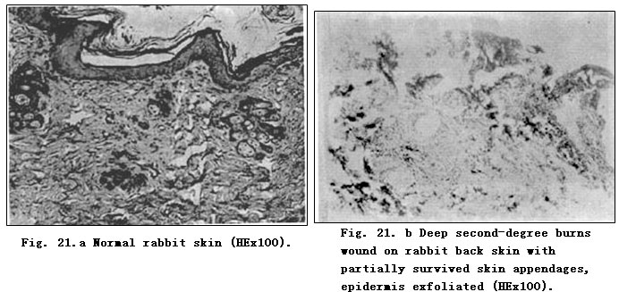
Normal rabbit skin demonstrated an absence of dermal papilla, but revealed abundant structures of skin hair and appendages (Fig. 21.a). Deep second-degree burns wounds on rabbit back skin involved deep dermis causing necrosis of full epidermis and partial dermis. Fibers in the dermis reticular layer and the subcutaneous layer appeared to be thick and sparse with partially survived skin appendages (Fig. 21. b). The results of pathomorphological examinations of two groups at different phases are shown in Table 56.
CONCLUSION
BRT with MEBT/MEBO treatment can make injured tissue regenerate in a relatively physiological environment that conforms to the natural law of tissue regeneration. As a result, scar formation is reduced to the maximum extent. These experimental results were in accordance with clinical observations.
DISCUSSION
Histologically, the regenerative capacity of skin tissue has a close correlation with tissue repair. Skin cells can be classified into two categories according to the different capability of regeneration: 1) Constantly changing cells,i.e. epidermal cells that have the ability to divide for indefinite periods under a normal state and proliferate to compensate shed and consumed cells. 2) Stable cells as epithelium in the skin body of a gland that cease proliferation when the organs mature, but have a continuous potential for division which is activated after injury to regenerate [1]. Tissue repair is also achieved by two approaches. First, by the regeneration of tissues similar both in structure and function- the structure and function of repaired tissue can be entirely identical to those of the original[2]. Take, for example, the tissue repair of superficial second-degree burns wounds. On days 3~4 postburn, epithelia began to grow, and continued thusly on days 5~8 and was mostly completed on days 8~10 postburn. Secondly, repair of the damaged tissues can be achieved through the formation of fibrous tissue, beginning with formation of granular tissues and ending with scar formation. Microscopic examinations of the tissue repair of deep second-degree and third-degree burns wounds showed an intertexture mainly comprised of fibroblasts and neoformative blood capillaries. Together with infiltration of plasmocytes (such as neutrophil, lymphocyte, plasma cell, macrophage), we note neoformative granulation tissues that were then replaced by a great quantity of closely aligned collagenous intercellular fibers. Subsequent to the decrease of fibroblasts, showing a long and narrow shape, and of blood capillaries, these tissues eventually developed into scars.
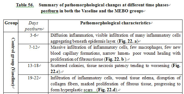
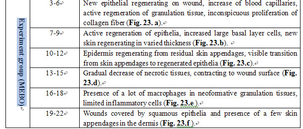
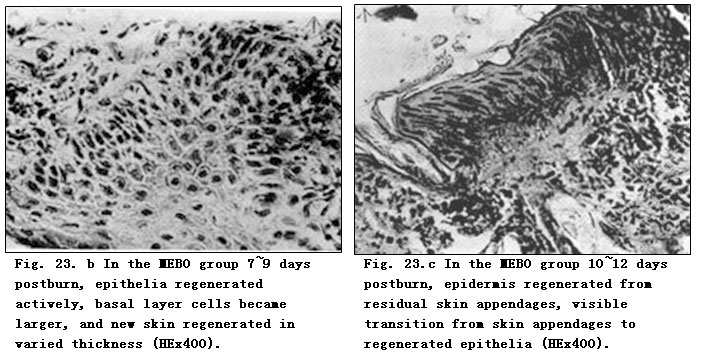
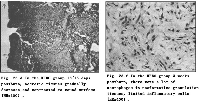
According to our experimental results, in the control group there was sparse blood capillary formation with narrow lumen. The tissue was swollen with fiber proliferation and massive infiltration of inflammatory cells. The disruption of collagen fiber and absence of regenerated epidermis to cover wounds eventually resulted in wound healing by eschar (showed in Fig. 22. d). In the experimental group treated with MEBO, residual skin appendages regenerated into epidermis with multilayers and large nuclei that progressed and covered the wounds (Fig. 23. b-d). Granulation tissue was promoted into regenerative tissue; neoformative blood capillaries were enhanced more than in the control group with larger lumen and richer blood supply, both of which facilitate an enhanced metabolism (Fig. 23. a). Finally, wounds in the experimental group were covered by squamous epithelia and healed without scarring (Fig. 23.g). We noted that when burns wounds were treated by BRT with MEBT/MEBO, the perpetually changing cells began to divide and proliferate toward the wound center along the wound edges or the basal part of residual epithelia, whereas after burns injury, the stable cells residing in skin appendages and grandular epithelium were activated to divide and regenerate into epidermic tissue that in turn migrated toward and finally closed the wound. Pathological examination on days 10-12 postburn showed a transitional migration from skin appendages to regenerated epithelia. When epithelia proliferated and divided, wounds were covered by stratified squamous epithelium. The macroscopic appearance of healed wounds was initially in red or pink and progressively became normal in color.
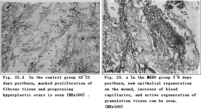

Wounds in the control group (Vaseline) showed slow repairing, obvious proliferation of fibrous tissue and healed with hyperplastic scars. By comparison, wounds in the experimental group (MEBO) expressed rapid repairing, active growth of neoformative epidermis, inconspicuous proliferation of fibrous tissue and eventually healed without scarring. These results demonstrate that MEBO retains optimal wound moisture, while tissue is not immersed. MEBO created a drug membrane that protected and isolated wound tissue from outer contaminant, allowing native histocytes to propagate in a relatively physiological environment in accordance with the nature regenerative law of skin. Local microcirculation was also improved and pathological changes of three zones of burns wounds (necrosis zone, stasis zone and hyperemia zone) were reversed. These conditions were favorable to the recovery of tissues in the stasis zone. Therefore, MEBO was believed to promote epithelial regeneration, control the increased speed of connective tissue, and keep epithelia and connective tissue in an almost normal rate of proliferation so as to heal deep burns wounds with less or minimal scarring.
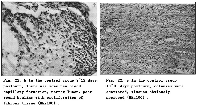
In the experiment, wounds were contaminated by S. aureus. Microscopic observation showed massive infiltration and aggregation of inflammatory cells in the Vaseline group with few macrophage and scattered colonies without boundaries (Fig. 22.c). All wounds were visibly infected within 1 week. In the MEBO group, inflammatory cells were large in quantity with enhanced capacity of anti-infection (Fig. 23.e). Gross observation revealed that wounds in this group repaired rapidly with absence of inflammatory response such as red swelling. It was believed that MEBO demonstrated efficacy in promoting blood circulation by removing blood stasis, clearing away heat and toxic material, relieving inflammation and removing the necrotic tissue while promoting granulation. The experiment also demonstrated that MEBO might inhibit or kill the growth of S. aureus.
In this study, the rabbit burns model was kept stable with zero mortality. Light-microscopic examination revealed a distinct process of histocyte repair. The results showed that the application of BRT with MEBT/MEBO in burns management could prevent and control infection, promote wound repair, minimize scar formation, shorten healing time, avoid complications and relieve pain as well. BRT with MEBT/MEBO also has the advantages of facilitating the observation of wound repair and easy application. BRT with MEBT/MEBO is now irrefutably considered to be the standard of care for burns management worldwide.
REFERENCES
1. Zhang YM: Experiences in treating facial scars by the combination of abrasive technique and excision. Proceedings of Cosmetic Symposium, Wuhan, 1990, p 142.
2. Wuhan Medical College (ed): Pathology, ed 1. Beijing, People’s Health Press, 1982, pp 19~21.
3. Academic Committee of the First National Conference of Moist Exposed Burn Therapy: A great historical turn in the burn medical science. Chin J Burns Wounds Ulcers 1989; 1: 4~10.