 企业邮箱
企业邮箱


Electron-Microscopic Observation of One Case of Skins Burns
作者:Rong Xiang Xu 出版社:KARGER 发行日期:In 2004INTRODUCTION
To further investigate the mechanism of deep burns wounds healing without hyperplasic scar formation after treatment with BRT with MEBT/MEBO, we took a biopsy from a deep wound site of a severely burned child before and after treatment in order to observe it via light and transmission electron microscope. The aim of the study was to find the histological evidence of scar-free healing.
CASE REPORT
A 12-year-old boy was admitted (PID. 172650) on November 4th 1989 after suffering direct gas flame burns on face, trunk and extremities. Clinical assessment indicated a total burns surface area (TBSA) of 75%, including 45% second-degree and 30% third-degree wounds. The condition of the patient remained stable during anti-shock therapy, but he developed sepsis on day 6 postburn. Serial blood cultures x 3 were negative. On day 10 postburn, escharectomy and microskin grafting were performed on the left upper and right lower extremities. On day 20, excision and microskin grafting were performed on the back again. On day 30, burns wounds of the right leg and dorsum pedis with mixed second- and third-degree as well as deep second-degree burns on the back were treated with BRT with MEBT/MEBO. Wound tissue biopsy was taken from the right leg before and after treatment, then pathological examinations were carried out light and transmission electron microscopically.
RESULT
Pathological examination revealed satisfactory healing of the burns wounds treated with MEBO without formation of obvious hyperplasic scar tissue.
1) Light microscopy
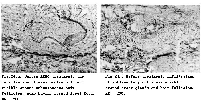
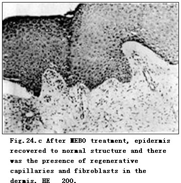
Before treatment, the infiltration of inflammatory cells was visible around sweat glands and hair follicles, some having formed local foci (Fig. 24.a, b). After treatment, skin recovered to normal structure with regenerative capillaries and fibroblasts in dermis (Fig. 24.c).
2) Transmission electron microscopic observation
Before treatment, a lot of circular vacuoles were present in the surrounding nucleus that showed irregular nuclear membrane with disappearance of nucleolus. Elastic fibers in the dermis varied thickness and had a disorderly arrangement with deposit in the lumen (Fig.25.c). After treatment, cells in the stratum spinosum became regular, showing distinct nucleus, clear nucleolus and uniform distribution of nuclear chromatin. Desmosome of the intercellular bridge recovered to normal (Fig.26.a-c).
CONCLUSION
The results proved that after using MEBO, the burns wounds healed without formation of macroscopic hyperplasic scar. The ultrastructure of the healing burns wound was similar to that of an ordinary traumatic wound.
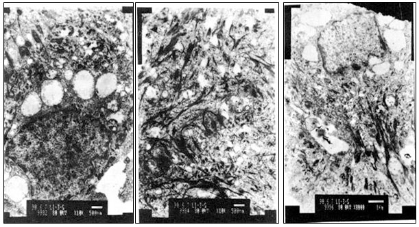
Fig.25.a (left). Before MEBO treatment, many circular vacuoles were present in the surrounding nucleus that showed irregular nuclear membrane with disappearance of nucleolus. TEM. 10,000.
Fig.25.b (middle). Before treatment, elastic fibers in dermis were in varied thickness and disorderly arrangement with vacuolar degeneration. TEM. 10,000.
Fig.25.c(right). Before MEBO treatment, appearance of irregular nucleus, presence of perinuclear vacuoles and disordered elastic fibers with deposit. TEM. 8,000.
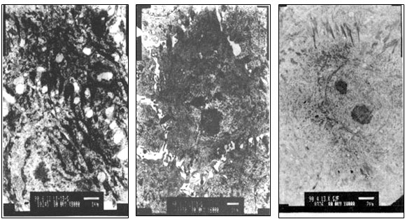
Fig.26.a (left). After MEBO treatment, intercellular bridge of cells in stratum spinosum recovered to normal with distinct nucleus and clear nucleolus. TEM. 4,000.
Fig.26. b (middle). After treatment, desmosome of cell junction almost recovered to normal with clear shape of cell, regular nucleus and uniform distribution of euchromatin. TEM. 6,000.
Fig.26. c (right). After MEBO treatment, structure of desmosome recovered to normal with uniform distribution of nuclear chromatin and regular cell shape with nucleolus in center. TEM. 5,000.
DISCUSSION
In dermis of normal skin, the dominant cell relating to traumatic repair and proliferative inflammation is the fibroblast. It is located adjacent to a collagenous fiber bundle, showing as fusiform, stellar or polygonal shapes, and having thick and short cell process. The fibroblast contains an oval nucleus which occupies one third of whole cell. It also reveals an obvious nuclear membrane and one or two nucleoli. There is expanded lumen of intracytoplasmic rough endoplasmic reticulum (RER). There are four major types of cell junctions between epithelial cells, i.e. tight junction, intermediate junction, gap junction and desmosome.
This study showed that perinuclear vacuoles, disordered elastic and collagenous fibers were presented before MEBO treatment, comparing to desmosome of intercellular bridge recovering to normal structure after MEBO treatment. When in the hyperfunction stage, the intracytoplasmic RER appeared as small fragmental vesiculiform, and when in vigorous synthesization, it appeared tight with flocculation within the cistern. The main function of the RER is to synthesize protein. Smooth endoplasmic reticulum (SER) has functions correlating with the synthesis of lipoids and steroids.
Stephen [1] reported the presence of myofibroblasts in hypertrophic scar tissue according to electron-microscopic observation. Myofibroblast contained incomplete nuclear membrane with developed RER. As it has both the characteristics of smooth muscle cells and the shape of fibroblasts, it is also termed “modified myofibroblast”. We have reported the ultrastructure of scar resulting from burns injuries in 1985 [2]. Comparison of the previous and present studies indicate that no macroscopic hypertrophic scar was formed on burns wounds treated with BRT with MEBT/MEBO, and the ultrastructure of the healing burns wound appeared no different from the ordinary traumatic wound. Though we have previously reported the clinical experience of applying MEBO for treating burns wounds of varying degrees [3], this was our first presentation of light-microscopic and transmission electron-microscopic observations regarding burns wounds. We would like to disclose our research achievement here in order to stimulate further studies.
REFERENCES
1. Stephen A: Wound contraction and fibrocontractive disorders. Arch Surg 1978; 1: 1034-1046.
2. Hong ST, et al: Ultrastructure of scars resulted from burns. Metal Med 1985; 1: 5-8.
3. Chen SR, Wang Y, Zhang XZ, et al: Clinical observation of the effect of moist exposed burn ointment (MEBO) on treating one case with extensive burn. Chin J Burns Wounds Surface Ulcers 1989; 1: 46.