 企业邮箱
企业邮箱


Clinical Procedure and Histological Observation of Full-Thic
作者:Rong Xiang Xu 出版社:KARGER 发行日期:In 2004INTRODUCTION
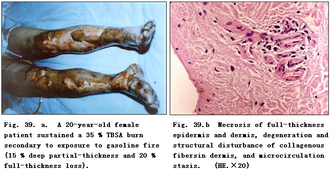
A 20 year-old female patient sustained a 35% TBSA burn secondary to exposure to gasoline fire (15% deep partial-thickness and 20% full-thickness loss). The patient was hospitalized at 12 h postburn and was diagnosed as suffering with full-thickness loss (III-degree) burns on both lower extremities. The epidermis was necrotic and detached and the dermal layer degenerated and necrotic with a waxy yellow and waxy white appearance (Fig.39.a). The pathological section examination of the sampled local wound tissues revealed necrosis of full-thickness epidermis and dermis, degeneration and structural disturbance of collagenous fibers in dermis, and microcirculation stasis (Fig.39. b).
RESULTS
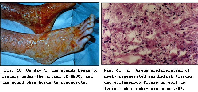
After admission, the patient was treated initially with MEBO burns ointment to protect burn tissue before we performed skin cultivation and relief as per our BRT protocol. At 48 h postburn, the wounds began to liquefy and the liquefaction was complete by day 4 (Fig. 40). The liquefied products were gently removed from the wound surface before MEBO was reapplied every 3~4 h.
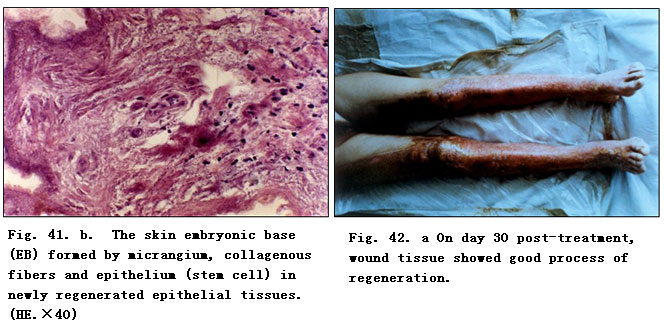
Repeat biopsy at the same burned location was performed for pathological examination. The results showed massive granular tissues among the necrotic epithelial tissue,a proliferation of newly regenerated epithelial cells with collagenous fibers, as well as the typical skin embryonic base (EB, Fig. 41. a,b). After a 10-day application of MEBO, the comparably primitive epithelial tissues were observed under pathological examination of epithelial tissues sampled from the wound edge.
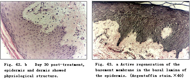
At 20 days post-treatment with MEBO, the pathological examination of deep burns wounds tissue showed the presence of the newly formed intact stratified squamous epithelium. The epithelial cells of the superficial layer appeared normal. The appearance of the collencytes and microangium in the dermis layer was typical. On day 30 post-treatment, epithelial tissues showed a remarkable degree of regeneration (Fig. 42. a), and skin structure was almost normal (Fig. 42. b).
Immunohistochemical Examination:
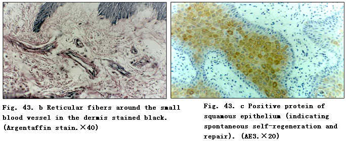
Twenty days post-treatment with BRT with MEBT/MEBO, wound tissue was examined and the results showed the clear appearance of collagenous fibers in epithelial tissue and subcutaneous tissue. Argentaffin stain indicated active regeneration of reticular fibers. The reticular fibers around the small blood vessels of subcutaneous tissue appeared relatively normal. The basal cells of epidermis regenerated actively (Fig. 43. a,b).
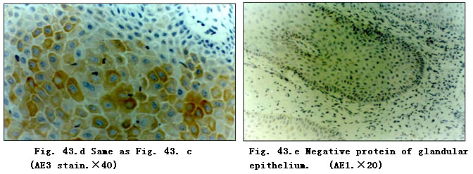
Thirty days after BRT with MEBT/MEBO treatment, the regenerated and repaired epithelial tissue was AE3 stained. The pictures showed positive proteins of squamous epithelium, brown-stained cytoplasm and blue-stained nucleus of granular cells in epidermis (Fig. 43. c,d).
AE1 stain showed negative proteins of glandular epithelium, which is representative of glandular epithelium already transformed into squamous epithelium (Fig. 43. e).
Electron-Microscope Observation:
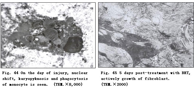
On the day of injury, the epithelium showed necrotic degeneration; and the monocytes showed a clear nuclear shift with karyopyknosis and even phagocytosis (Fig. 44) .
Five days post-treatment with MEBO, we noted active growth of fibrocytes and fibroblasts (Fig. 45).
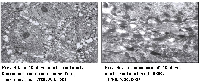
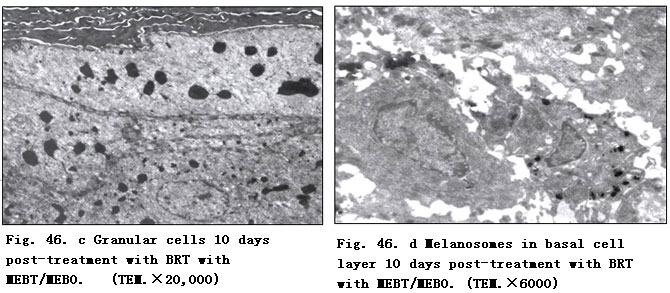
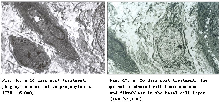
Ten days post-treatment with BRT with MEBT/MEBO, we noted the appearance in epidermis of echinocytes and desmosome in the stratum spinosum (Fig. 46. a, b), granular cells in stratum granulosum, and melanosomes in the basal cell layer (Fig. 46. c, d). We also observed phagocytes active around the small vessels, indicating recovery of function (Fig. 46. e).
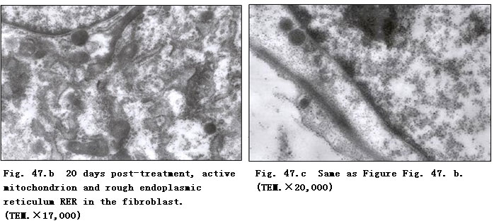
Twenty days post-treated, we noted the appearance of the hemidesmosome junction between the basal cell layer and epithelia. Active mitochondria and RER in fibroblasts also appeared (Fig. 47. a-c).
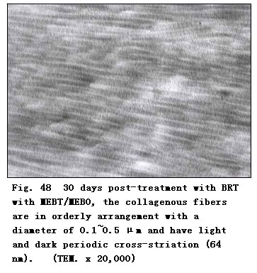
Thirty days post-treatment with BRT with MEBT/MEBO, with the regeneration and repairing of epithelium almost complete, collagenous fibers were mature with a diameter of 0.1~0.5 μm and arranged in an orderly fashion (Fig. 48). Light and dark periodic cross-striation (64 nm) was also observed. No pathomorphological changes of collagenous fibers such as distortion, helicoid (whirlpool) or cauliflower-like were observed.
After wound healing, functional exercises and physiotherapy of the lower extremities were required. MEBO was continued as ordinary skin oil. The patient healed and was discharged to home on day f45 postburn.