 企业邮箱
企业邮箱


世界生命科学前沿动态周报(九十)
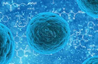
培养生成的肝脏干细胞被移植后显示治疗效果
2012年2月25日科技日报:由于肝细胞具有丰富的生物医学作用,如用于肝炎、药物代谢和毒性、肝硬化移植以及其它慢性肝病的研究,数十年来,世界各地的科学家一直试图再生出原代肝细胞。可到目前为止,也没有实验室能够使用任何可用技术,在培养液中成功鉴别和培育出肝脏干细胞。
在近期的《自然》杂志中,美国俄勒冈州波特兰市朵贝克儿童医院俄勒冈州健康科学大学佩普家庭儿科研究所内科科学家们和荷兰乌特勒支Hubrecht发展生理学和干细胞研究所的研究员们描述了一种能够在培养皿中无限扩增小鼠肝脏干细胞的新方法。
“此研究为以类似方法培育出与小鼠肝脏干细胞相似的人类肝细胞,并有效地转化成功能性肝细胞,带来了希望,” Markus Grompe 博士说。他是该研究的研究成员,美国俄勒冈州波特兰市朵贝克儿童医院俄勒冈州健康科学大学佩普家庭儿科研究所所长,同时也是该大学医学院儿科、分子与医学遗传学教授。
在《自然》之前刊登的一篇研究中,由Hubrecht研究所Hans Clever医学博士领导的研究员们,通过观察成人干细胞标记物Lgr5的表达及其在生长因子Wnt作用下的生长情况,首次在小肠和结肠中鉴别出了干细胞。他们猜测Lgr5的独特表达类型也可用于标记其他成体组织干细胞,如肝脏干细胞;之前如何确认肝脏的干细胞一直是无从得知的事情。
在本期《自然》刊登的研究中,Grompe和其同事们对Clever博士的研究方法进行了改进,结果发现Wnt诱导的Lgr5表达不仅可以标记肝脏干细胞的生成,还可标记肝脏受损时变活跃的一种干细胞。
科学家们能够在培养皿中成倍地生成肝脏干细胞,这是前所未有的成果。随后这些生成的干细胞被移植到一种专门设计的肝病小鼠模型中,发现它们依然可以生长并表现出一定的治疗效果。
“我们能够大量生成肝脏细胞并把它们以适度的比例转化成肝实质细胞。接下来,我们将利用其它的生长因子和条件来提高这个转化百分比。人类慢性肝脏疾病的肝脏干细胞疗法即将来临,”Grompe 博士说。
【点评】该研究在培养皿中成功生成肝脏干细胞并在肝病小鼠模型中实现动物临床试验治疗效果,对干细胞研究,特别是肝脏干细胞研究具有很大地促进作用,不过就此推测人类肝脏慢性病的干细胞疗法即将来临,还为时尚早。这种体外培植的干细胞能不能规避移植后的排斥反应这一难题,仍需做大量的临床研究工作。
相关文献:http://www.nature.com/nature/journal/v494/n7436/full/nature11826.html
In vitro expansion of single Lgr5+ liver stem cells induced by Wnt-driven regeneration
Authors: Meritxell Huch, Craig Dorrell, Sylvia F. Boj, Johan H. van Es, Vivian S. W. Li, Marc van de Wetering, Toshiro Sato, Karien Hamer, Nobuo Sasaki, Milton J. Finegold, Annelise Haft, Robert G. Vries, Markus Grompe & Hans Clevers
The Wnt target gene Lgr5 (leucine-rich-repeat-containing G-protein-coupled receptor 5) marks actively dividing stem cells in Wnt-driven, self-renewing tissues such as small intestine and colon1, stomach2 and hair follicles3. A three-dimensional culture system allows long-term clonal expansion of single Lgr5+ stem cells into transplantable organoids (budding cysts) that retain many characteristics of the original epithelial architecture2, 4, 5. A crucial component of the culture medium is the Wnt agonist RSPO16, the recently discovered ligand of LGR57, 8. Here we show that Lgr5-lacZ is not expressed in healthy adult liver, however, small Lgr5-LacZ+ cells appear near bile ducts upon damage, coinciding with robust activation of Wnt signalling. As shown by mouse lineage tracing using a new Lgr5-IRES-creERT2 knock-in allele, damage-induced Lgr5+ cells generate hepatocytes and bile ducts in vivo. Single Lgr5+ cells from damaged mouse liver can be clonally expanded as organoids in Rspo1-based culture medium over several months. Such clonal organoids can be induced to differentiate in vitro and to generate functional hepatocytes upon transplantation into Fah−/− mice. These findings indicate that previous observations concerning Lgr5+ stem cells in actively self-renewing tissues can also be extended to damage-induced stem cells in a tissue with a low rate of spontaneous proliferation.
Subject terms:
• Stem cells